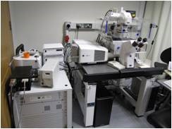
Cell Dive Spatial Proteomics
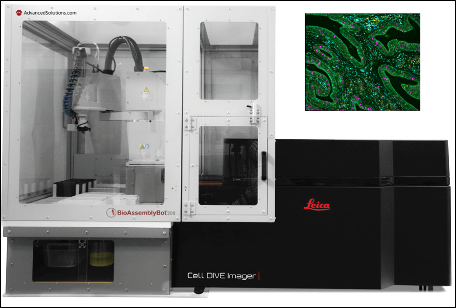
Purpose of this equipment: Spatial proteomics analysis of fixed samples, with a resolution down to below single cell level with access to ca. 60 targets. Samples can be stained with up to 4 different fluorescent markers plus Hoechst/DAPI is used as alignment aid for sequential imaging steps (Antibodies can be imaged using 470, 542, 631 and 730 nm excitation with 20-40nm band width, a 5 band multi chroic and 5 single band pass emission filters. A whole sample overview is given and will be used to select regions of interest manually or automatically.
External academic users costs: Training/Supervision – €150/hr. Unsupervised sessions – €74/hr. Service €150/hr. This does not include consumables. Data will be stored for 1 week on the instruments workstation. Users should provide external networked data storage. Contact hduessmann@rcsi.ie for more details of the instruments capabilities.
Nanostring GeoMX DSP for single cell transcriptomics and proteomics
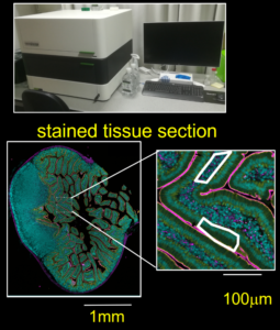
Purpose of this equipment: Spatial transcriptomics and proteomics analysis of fixed samples, with a resolution down to below single cell level with access to e.g. ~20k targets for whole transcriptome studies. Samples can be stained with up to 4 different fluorescent markers (488, 532, 594 ann 633 nm excitation, whole sample will then be scanned allowing to select regions of interest manually or automatically.
External academic users costs: Training/Supervision – €150/hr. Unsupervised sessions – €60/hr. Service €150/hr. This does not include consumables. Data will be stored for 1 week on the instrument, users should provide external networked data storage
Contact iwoods@rcsi.ie for sample preparation and bookings:
contact hduessmann@rcsi.ie for more details of the instruments fluorescence imaging capabilities and data handling.
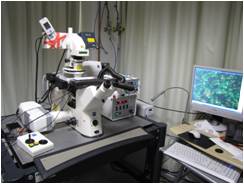
Zeiss PALM Microbeam Fluorescence and transmitted light Microscope with live cell imaging capability.
Purpose of this equipment: laser capture micro-dissection from live or fixed samples, live cell imaging, coordinate transfer for sample retrieval between LSM 980 and PALM Microbeam.
Inverted microscope, small stage incubator, hardware autofocus, laser lines: 355nm for laser micro-dissection and capture.
External academic users costs: Training/Supervision – €150/hr. Unsupervised sessions – €55 half day, €55 whole night. Service €90/hr. Incubation €5/time-slot. This does not include consumables. Data will be stored for a minimum of 1 week on the joint image processing workstation. Users should provide external networked data storage. Contact hduessmann@rcsi.ie for more details of the instruments fluorescence imaging capabilities, for support and bookings
Zeiss LSM 980 Airyscan 2 Confocal Microscope with super resolution capability and TCSPC by Becker and Hickl.
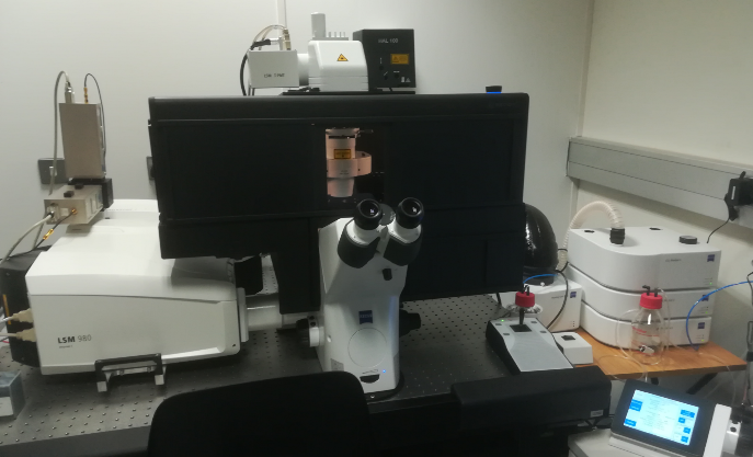
Purpose of this equipment: live cell imaging, super-resolution imaging on live and fixed samples, FRET incl FLIM-FRET, 3D imaging, FRAP, PLIM. Inverted microscope, large stage incubator, z-piezo stage, hardware autofocus, laser lines: 405, 445, 488, 514, 561, 639 nm. For FLIM, PLIM and FCS: 405 and 488 nm pulsed lasers.
External academic users costs: Training/Supervision – €150/hr. Unsupervised sessions – €91 half day, €91 whole night. Service €200/hr. Incubation €5 per time-slot. This does not include consumables. Data will be stored for a minimum of 1 week on the joint image processing workstation. VUsers should provide external networked data storage. Contact hduessmann@rcsi.ie for support and bookings.
Nikon TE2000, Automated Epi-fluorescence Microscope
Purpose of this equipment: Live cell imaging, FRET, multiple fields of view imaging, Autofluorescence (NADH), multiple fields of view.
Information: Inverted microscope, small stage incubator, excitation and emission filter wheels, motorised stage, back illuminated EM-CCD (Andor ixon DU-897), MetaMorph
External academic users costs: Training/Supervision – €150/hr. Unsupervised sessions – €20 half day, €20 whole night. Service €120/hr. Incubation €5.00/time-slot
Contact hduessmann@rcsi.ie for support and bookings.
Light Sheet Fluorescence Microscope LSFM (Lightsheet Z1).
The LSFM is suitable for live biological samples using a 20x 1.0 NA water immersion imaging lens and 2x 5x 0.1NA cylindrical lenses as well as optically cleared samples using a dedicated 20x immersion objective with 1.0NA and 2 x 10x 0.2NA Cylindrical lenses. Excitation at 405, 488, 561 and 638 nm is available. For booking and costs OR a description of spectral properties of the imaging light path please contact hduessmann@rcsi.ie
see link below for two examples – Using the clearing techniques: Nice video of a view into a hippocampus of tie-2-GFP expressing mouse. Clearing, imaging and image processing (FiJi, Blender, Fluorender) by Brenton Cavanagh using the Lightsheet Z1.
N14-Spheroid2-Hoechst-PI-Fusion Of 5 Dual Views: Dual label live cell imaging of Nuclei in a N14-1208 patient-derived glioblastoma neurosphere with Hoechst (cyan) and PI(red, stains only dead cells), sample preparation by Viktorija Juric, imaging and image processing (FiJi) by Heiko Düssmann using the Lightsheet Z1.
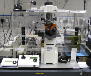
AIS-2: Automated microinjection system on Zeiss Aviovert 200M Epi-fluorescence Microscope
Purpose of this equipment: automated microinjection of multiple fields of view, live cell imaging, FRET
Information: inverted microscope, incubator, excitation and emission filter wheels, motorised stage, EM-CCD (Hamamatsu Orca 9100-02), AIS2 and Axiovision
External academic users costs: Training/Supervision – €150/hr. Unsupervised sessions – €30 half day, €30 whole night. Service €120/hr. Incubation €5.00/time-slot
Contact: Heiko Dussmann – email: hdussmann@rcsi.ie
Location: RCSI York Street, Dublin 2, Ireland
Zeiss LSM 710 Confocal Microscope.
Purpose of this equipment: live cell imaging, spectral imaging, FRET, 3D imaging, FRAP Inverted microscope, small stage incubator, piezo stage insert, hardware autofocus, laser lines: 405, 458, 488, 514, 543, 561, 633 nm. External academic users costs: Training/Supervision – €150/hr. Unsupervised sessions – €50 half day, €50 whole night. Service €150/hr. Incubation €5.00/time-slot. Contact hduessmann@rcsi.ie for support and bookings.
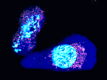
Nice example of a 3D reconstruction of live DU145 prostate cancer cells stained with Calcein (cytosol, dark blue), TMRM (functional mitochondria, red) and Hoechst (DNA, cyan), the left cell is dividing, acquired on LSM 710 by Heiko Düessmann.
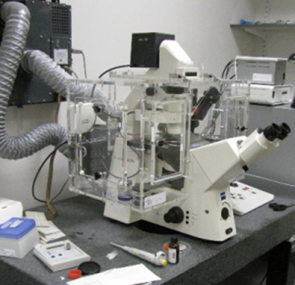
Zeiss LSM 5 live Duoscan Laser Scanning Microscope.
Purpose of this equipment: live cell imaging, FRET, fast 3D imaging, FRAP Information: INDIMO 1080, inverted microscope, small stage incubator, piezo focus, motorised stage, laser lines: 405, 489, 561 nm.
External academic users costs: Training/Supervision – €150/hr. Unsupervised sessions – €50 half day, €50 whole night. Service €150/hr. Incubation €5.00/time-slot
email hduessmann@rcsi.ie for support and booking information
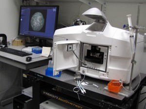
Cellomics Arrayscan VTI (including incubator & liquid handling robot)
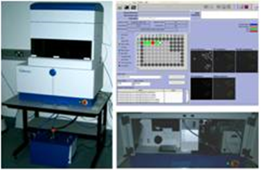
Purpose of this equipment: fluorescence imaging of large populations of cells in culture.
Information: inverted fluorescence wide-field microscope, dry lenses, motorized stage compatible with multiwell or slide formats, CO2 and temperature control, liquid handling system.
External academic users costs: Training/Supervision – €150/hr. Unsupervised sessions – €80 whole day. Service €150/hr. Incubation €5.00/time-slot
mail hduessmann@rcsi.ie for support and booking information
Zeiss Cell discoverer 7:
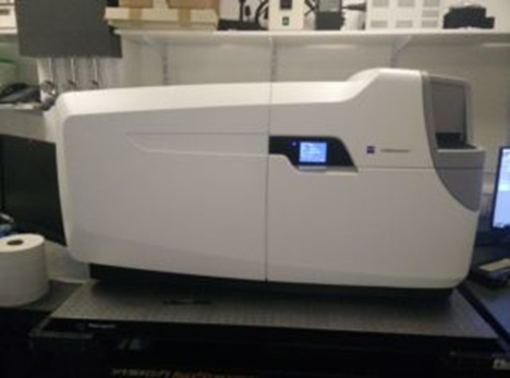
An automated boxed microscope system calibrates itself, detects and focuses on the samples while the optics adjust themselves. This makes it a reliable high content screening live cell imaging platform. Person to contact for training and advice on the CD7: Dr Brenton Cavanagh, Research Engineer for Microscopy, Office of Research and Innovation: email –brentoncavanagh@rcsi.ie for support and booking information.
Hitachi H7650 Transmission Electron Microscope
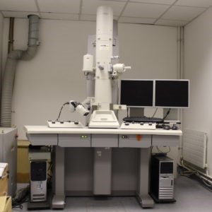
A high resolution microscope capable of resolving objects as small as 2nm. It allows the visualization of objects that are otherwise invisible to light microscopy. It is suitable for thin sections of biological sample, DNA origami and nanoparticle characterization. Person to contact for training and advice on the TEM: Dr Brenton Cavanagh, Research Engineer for Microscopy, Office of Research and Innovation: email brentoncavanagh@rcsi.ie for support and booking information.
Laser Capture Microdissection:
External academic users costs: Training/Supervision – €150/hr. Unsupervised sessions – €62 half day, €62 whole night. Service €150/hr. Incubation €5.00/time-slot
****** All pricing is subject to VAT and external users will have to provide proof for liability insurance.